Tags & Description
What makes up the cardiac silhouette?
heart
pericardium
pericardial fat
fluid
What is the abnormality?
Gas in the pericardial space
***How big should the heart big on lateral rads?
How much sternal contact?
Dogs: 2.5-3.5 ICS
Cats: 2-3 ICS
Sternal contact: 2.5-3 ICS
On a lateral radiograph, what structures summate from 9-11 o’clock?
main pulmonary artery/pulmonary trunk
aorta
right auricle
Where is the left atrium located on a lateral projection?
At the carina
Can sometimes see a backpack sign
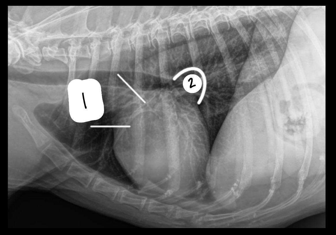
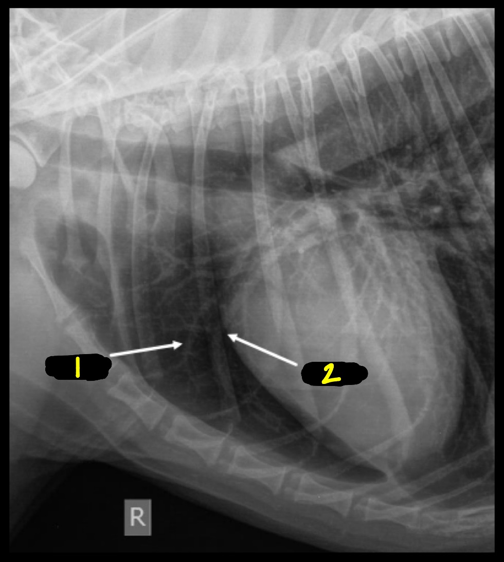
What is located at 1? At 2?
1 = right auricle, aorta, main pulmonary artery
2 = left atrium
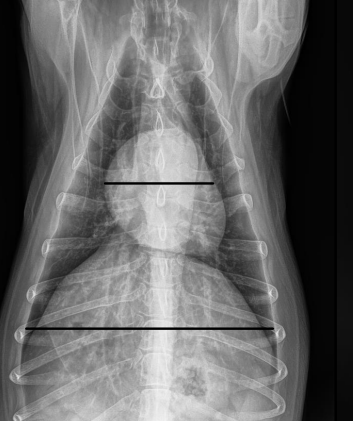
How large should the heart be on VD/DV view?
2/3 the width of the thoracic cavity
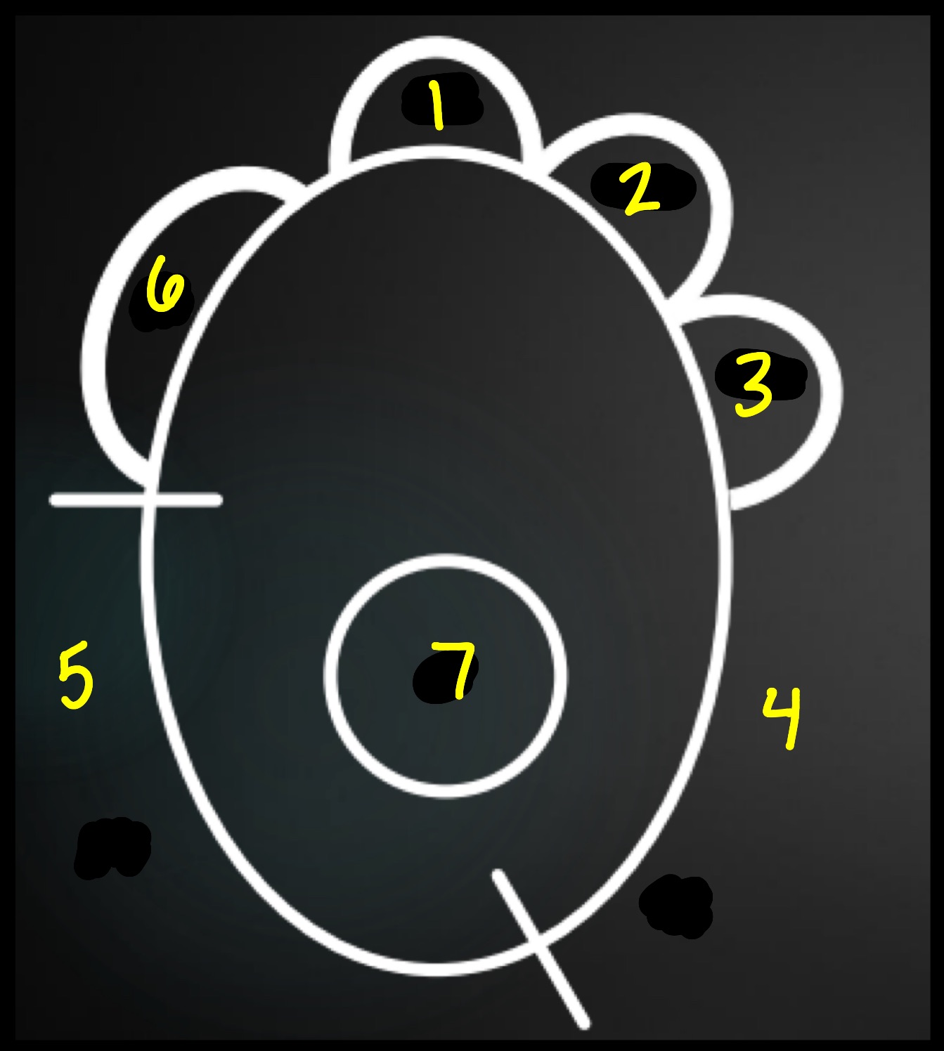
***What is the DV/DV clock face analogy?
11-1 o’clock = aortic arch
1-2 o’clock = main pulmonary artery
2-3 o’clock = left auricle
3-6 o’clock = left ventricle
6-9 o’clock = right ventricle
9-11 o’clock = right atrium
***What is 1-7 on this image?
1: aortic arch
2 = main pulmonary artery
3 = left auricle
4 = left ventricle
5 = right ventricle
6 = right atrium
7 = left atrium
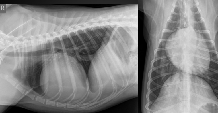
What are ddx for a globoid silhouette?
pericardial effusion
DCM
peritoneopericardial diaphragmatic hernia
tricuspid valve dysplasia
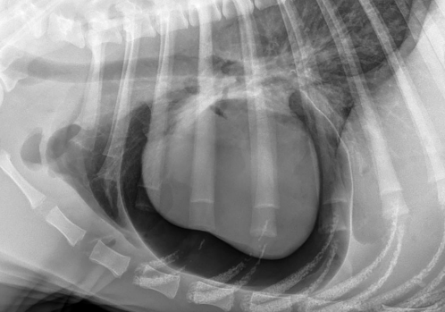
What is the shape of this heart? What are ddx?
Shape: globoid
pericardial effusion
DCM
peritoneopericardial diaphragmatic hernia
tricuspid valve dysplasia
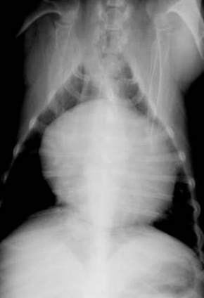
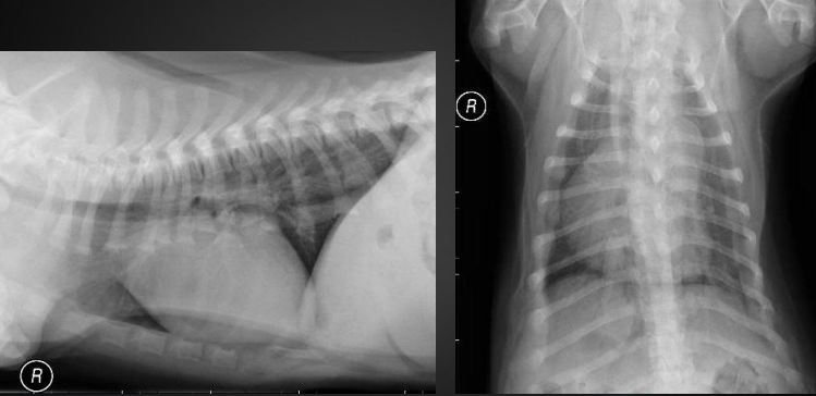
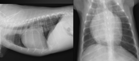
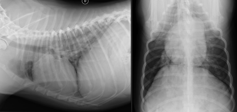
What is the diagnosis here?
Peritoneal pericardial diaphragmatic hernia
-not great diaphragm margins
-has structure in cardiac silhouette
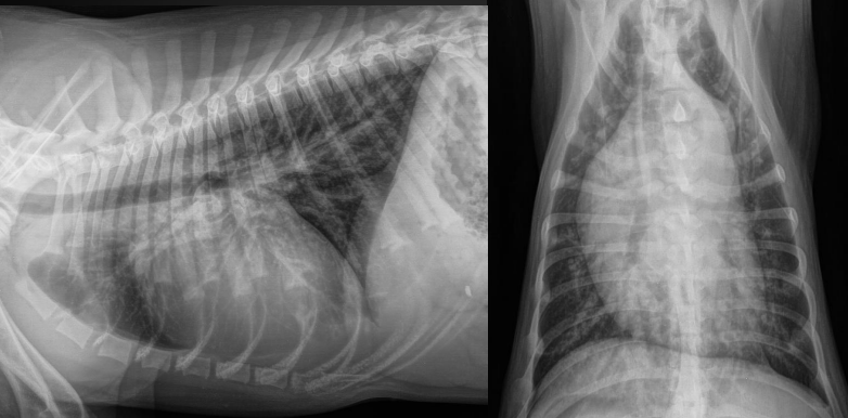
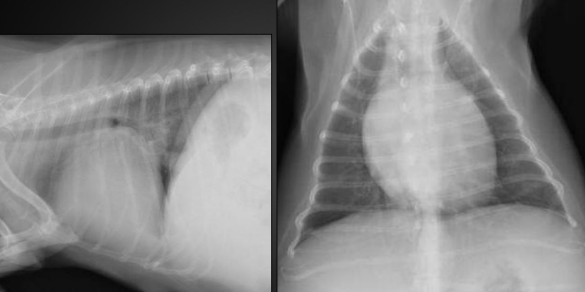
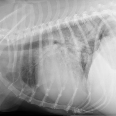
What is the heart shape?
Diagnosis?
Globoid
Pericardial effusion in this case
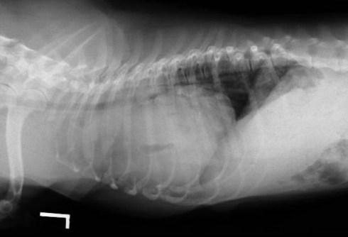
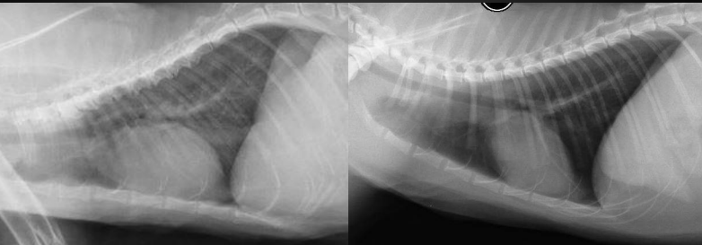
What are these examples of?
PPDH
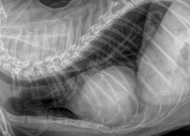
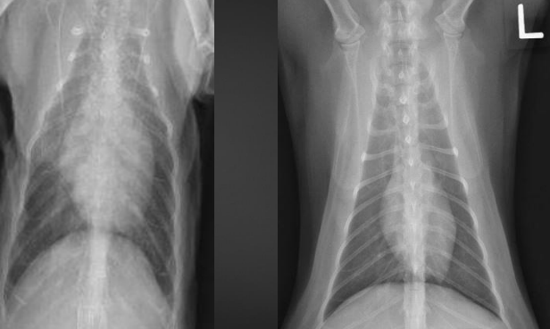
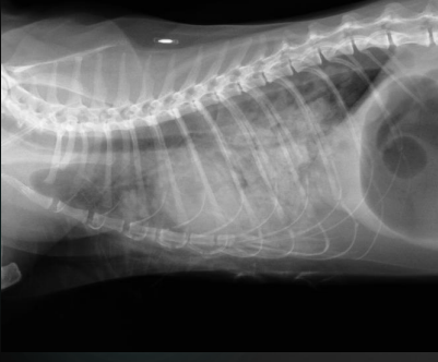
What is the diagnosis?
How do we know?
PPDH
region of increased ST opacity
possible stones in bile duct of liver
What is a normal vertebral heart scale score?
9.7 +/- 0.5 vertebrae, starting at T4
What is the vertebral heart scale useful for?
Monitoring
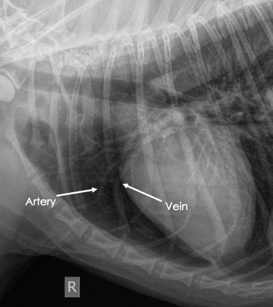
***How to remember which are veins and which are arteries?
Veins: ventral and central
Arteries: up and away
What is 1 and 2 in this image?
1 = artery
2 = vein
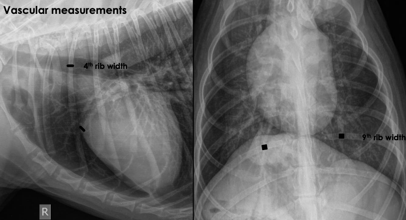
***How big should pulmonary arteries and veins be?
Should be same size
Lateral projection: cranial vessels
size of proximal 4th of 4th rib
VD projection: caudal vessels
size of 9th rib where they overlap
1.22 right caudal vein to 9th rib
(right caudal vein can be a little large than the 9th rib and still be normal)
****
What type of hypertrophy does volume overload cause?
Does this cause apparent cardiac enlargement?
Example?
eccentric hypertrophy
TIP: think eccentric person is outgoing, pushing out
apparent cardiac enlargement (large lumens)
shunting lesions
***
What type of hypertrophy does pressure overload cause?
Does this cause apparent cardiac enlargement?
Example?
concentric hypertrophy
thickened muscles, small lumens, not really outwardly apparent enlargement
stenotic lesions
***What are the 4 most common congenital cardiac diseases in small animals? What is most common in large animals?
Small animals
aortic stenosis
pulmonic stenosis
patent ductus arteriosus
tricuspid valve dysplasia
Large animals
ventricular septal defect
What are the radiographic findings?
Diagnosis?
bulge of aorta
11-1 o’clock on VD
can’t distinguish from aorta, pulmonary artery, or right auricle on lateral
Aortic stenosis
What are the radiographic findings?
Diagnosis?
bulge of main pulmonary artery
1-2 o’clock on DV/VD
can’t distinguish from aorta, pulmonary artery, or right auricle on lateral
What are the radiographic findings?
Diagnosis?
aorta bulge, MPA bulge
three knuckle appearance (aorta bulge, main pulmonary artery bulge, left auricle bulge) - not seen here, but common in PDA dogs
enlarged vessels
PDA
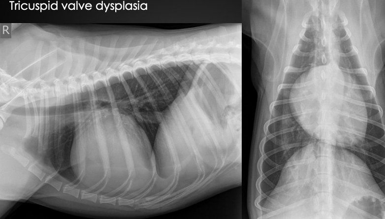
What are the radiographic findings?
Diagnosis?
Globoid cardiac silhouette
centered around right atrium (we don’t need to be that specific)
What are clinical signs/signalment of tricuspid valve dysplasia?
young dog
right sided murmur
***What are examples of acquired left-sided heart disease?
degenerative mitral valve disease (75% of cases)
DCM
may also cause right-sided disease
hypertrophic cardiomyopathy (cats)
***What are examples of acquired right-sided heart disease?
heartworms
tricuspid valve disease
can be acquired or congenital
acquired: can have right-sided changes as well if MVD present
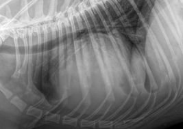
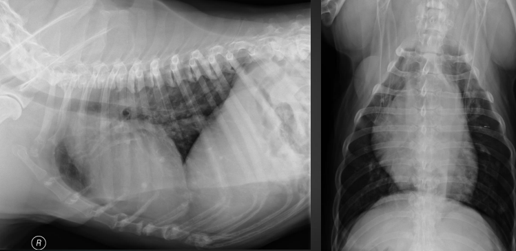
What are the radiographic findings?
Diagnosis?
Findings on lateral:
loss of caudal cardiac waist (flat line)
bulge of left atrium
dorsal deviation of trachea
Findings on DV/VD:
bulge at 2-3 o’clock = left auricle
enlarged left atrium (double wall sign)
a little right sided rounding
Diagnosis: mitral valve degeneration
probably some tricuspid valve degeneration as well b/c of right-sided rounding
What are the radiographic findings?
Diagnosis?
Findings:
loss of cardiac waist
rounding of right side
left atrial enlargement
Not definitive diagnosis until echo performed
likely DCM
What are the radiographic findings in this cat on the left? (normal on right)
Diagnosis?
Findings:
fat potato heart
Likely HCM
***Ddx for enlarged hearts in cats?
HCM (most common)
restrictive CM
thyrotoxic CM
dilated
unclassified CM
*Radiographs are not definitive for underlying cause for feline heart disease
*Echocardiogram needed to further investigate
What are the radiographic findings for the cat on the left? (normal on right)
Diagnosis?
Findings:
fat potato heart
left-sided enlargement
Diagnosis
HCM most likely
What are the radiographic findings?
Diagnosis?
Findings on VD:
pulmonary artery enlargement
bulge of MPA
reverse D
Findings on lateral:
possibly increased sternal contact (right ventricular enlargement)
Diagnosis: HW disease
***What are radiographic signs of left-sided heart failure?
cardiogenic pulmonary edema'
unstructured groundglass opacity, possibly consolidation
pulmonary venous congestion
left sided cardiac changes
atrial/ventricular enlargement
dorsal deviation of trachea
cats: can have pleural effusion
*Diuretics can cause vessels to be normal or small
***What are radiographic signs of right-sided heart failure?
distended CVC
hepatomegaly
peritoneal effusion
pleural effusion
right sided cardiac changes
rounding of atrioventricular region, possible bulge of main pulmonary artery depending on disease location
What are the radiographic findings in a cat?
Diagnosis?
Findings:
more ventral distribution of increased opacity
groundglass opacity
Diagnosis:
CHF
What are the radiographic findings in a dog?
Diagnosis?
Findings:
peri-hilar or caudodorsal ground-glass opacity
Diagnosis: CHF
What start of MVD do we start using medical mangement?
Stage B2 (left atrial or ventricular enlargement on radiograph)