


Nervous System Part 2-1
Electrochemical Activity
Activity in the neurons involves the movement of the following ions:
Sodium (Na +):
Found more on the outside of the neuron.
Chloride (Cl - ):
Found more on the outside of the neuron.
Potassium (K +):
Found more on the inside of the neuron.
large proteins ( A-):
Found more on the inside of the neuron.
The interiors of a neuron contain intercellular fluid.
The exterior of the neuron contains extracellular fluid.
Neuron Membrane

Electrochemical Activity
The semipermeable membrane:
Acts like a filter allowing the passage of some ions and preventing the passage of others.
During the resting state of the neuron, more positively charged potassium ions (K+) are actively moved to the interior of the neuron, and more positively charged sodium ions (Na+), and negatively charged chloride ions (Cl-) are actively moved to the exterior of the neuron.
The large proteins (A-) are part of the cell membrane and thus don’t move across it.
Proteins are trapped, creating a negative charge which then causes the inside to be negatively charged relative to the outside when in a resting state/resting potential.
Membrane Potential
membrane potential:
When the inside of a neuron has a negative charge compared with the outside.
Resting potential
The difference between the inside and outside of a neuron when it is not sending a signal is typically around -70mV.
The forces that maintain resting potential:
An uneven distribution of ions in both intracellular and extracellular parts of the neuron.
Two forces that cause this include the following:
Diffusion/concentration gradient
The movement from an area of high concentration down to an area of low concentration. (Na+ pulled in, Cl- pulled in, K+ pushed out.)
Electrostatic force
The interior of the neuron is negatively charged relative to the outside, which means positive ions are pulled in, and negative ions are pushed out. (Na+ pulled in, Cl- pushed out, K+ pulled in.)
In general, always remember that Na+K+ Pump.
The Sodium-Potassium Pump
The sodium-potassium pump does and is the reason for the following:
A process for actively pumping Na+ out of neurons and pumping K+ in.
Maintains polarized state/keeps equilibrium from forming.
Keeps the polarized state necessary to get nerve impulses.
If there weren’t a maintained polarized state, we would not get nerve impulses.
If there is a poisoning of the pump, there won’t be any nerve impulses.
If you eat a puffer fish that has its toxin still inside it (Tetrodotoxin), your nerve impulses would be blocked, and the result can be fatal.
Depolarization, Hyperpolarization & Repolarization
Depolarization:
When Na+ enters the neuron & it makes less of a negative charge on the inside, making it more likely to reach the threshold and achieve action potential/fire.
Hyperpolarization:
A state that is more negative than the resting state which makes it less likely to achieve action potential.
Repolarization:
The return of the resting state.
Activity Inside Dendrites
Na+ moves inside dendrites, and the inside of the dendrites become less negatively charged, also known as depolarized/depolarization.
As the dendrite moves down, the amount of Na+ moving in is decreased, which is known as decremental.
If the dendrites have enough Na+ moving in to achieve -55mV at the axon hillock, it will reach the threshold and achieve action potential/nerve impulse.
The Threshold of a Neuron
The threshold of a neuron occurs at the axon hillock.
Although different neurons have different thresholds, the most common threshold is often -55mV.
Action Potential (AP)
Also known as nerve impulse.
Occurs inside the axon.
A brief wave of positive electrical charges moves down the axon.
A wave of positive ions that are moving in is called Depolarization.
Inside becomes positive relative to the outside (inside at +30 mV or +40 mV)
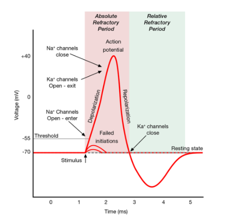

Action Potential (AP) propagates down the axon/follows a domino effect
sodium moves in at one part of the axon, causing the voltage-dependent gate to open in the next part of the axon, Na+ moves in, and so on down the axon.
Non-decremental
Does not decrease in intensity as it moves down the axon; the same amount of Na+ that is moving in is the same as it moves down.
When Action potential (AP) gets to the terminal buttons, it causes neurotransmitters to be released into the cleft.
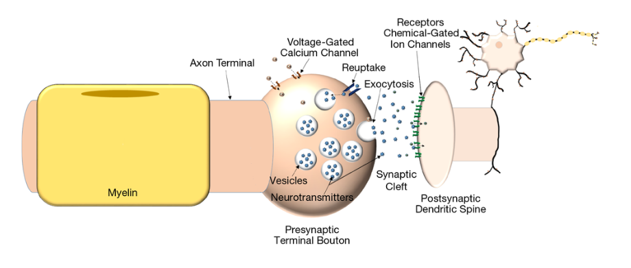
Postsynaptic Potentials
Neurotransmitters plug into receptor sites on the postsynaptic membrane and can cause one of the following:
EPSP: Excitatory postsynaptic potential:
Excitatory: increases the likelihood of the postsynaptic
neuron firing.Na+ moves in, Making it less negative inside.
Causes depolarization.
IPSP: Inhibitory postsynaptic potential:
Inhibitory: reduces the likelihood of the postsynaptic
neuron from firing.Cl- moves in: makes a more negative charge on the
inside.Causes hyperpolarization.
Refractory Periods
Refractory periods are also known as times when the neuron can’t fire.
Absolute:
Doesn’t fire, no matter how strong the stimulus is.
Relative:
Takes more substantial amounts of stimulus but can achieve action potential/ fire if enough stimulation occurs.
Absolute refractory period:
1-2 milliseconds during an action potential when another action potential cannot be produced
Relative refractory period:
The short time when the neuron membrane is hyperpolarized (more negative than resting potential.)
Difficult but not impossible to generate another action potential, but will need a powerful stimulus.
Graded Potentials Vs. Action Potentials
The graded potential is also known as activity inside dendrites.
Graded potential affects whether there will be enough activity at the axon hillock to reach the threshold & fire neurons.
Many messages come into dendrites from other neurons.
Whether there is enough activity to reach the threshold is determined by the additive effects of the messages called “Summation.”
Summation
There are two types of Summation, those being:
Temporal: 2 EPSPs come close together in time at the same place on dendrites or a cell body.
Spatial: 2 EPSPs arriving at different places on dendrites or a cell body and combine.
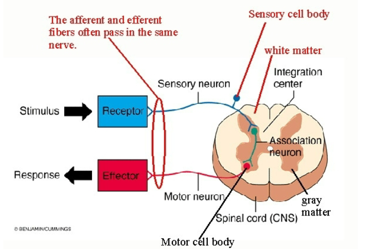
Fibers & Cell Bodies
Nerves:
Groups of fibers in the PNS (axons).
Tracts:
Groups of fibers in the CNS (axons).
Ganglion:
(Ganglia) cell bodies in the PNS.
Nucleus:
(Nuclei) cell bodies in the CNS.
Gray matter:
cell bodies
White matter:
Axons (myelinated)
Don’t confuse nerves with neurons; nerves are bundles of axons in the peripheral nervous system and are much larger structures than a neuron.
Types of Nerves: Reflex Arc
Afferent or Sensory Nerves:
Bundles of sensory fibers that carry sensory information into the spinal cord.
Motor Efferent Nerves:
Bundles of motor fibers that carry motor commands & messages from the spinal cord to muscles.
Mixed Nerves:
Bundles of sensory & motor fibers.
Example
Touch something hot: Sensory & Motor fibers
travel together in mixed nerves up the arm.As it gets to the spinal cord, Sensory nerves branch
off and enter the spinal cord through the back (dorsal
root.)Want to pull your hand away: Motor nerves carry
message out to muscles; come out from the front
(ventral root.)
Other Structures & Types of Cells in the Nervous System
Meninges:
Coverings of the brain & spinal cord.
Meningitis:
Infection of meninges
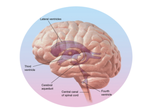
Central canal:
Runs the length of the spinal cord filled with CSF (cerebrospinal fluid.)
Ventricles:
Fluid-filled cavities/chambers in the brain; continuous with central canal Filled with CSF.
Hydrocephalus:
The buildup of fluid in ventricles; Ventricles enlarge, putting pressure on the brain
Treatment: shunt to drain fluid.
Functions of the Glial Cells include:
Provide nutrients
Create myelin
Remove dead neurons
Repair
Protect blood-brain barrier
