


Chapter 21: The Lymphatic and Immune System
Anatomy of the Lymphatic and Immune Systems
The immune system is the complex collection of cells and organs that destroys or neutralizes pathogens that would otherwise cause disease or death.
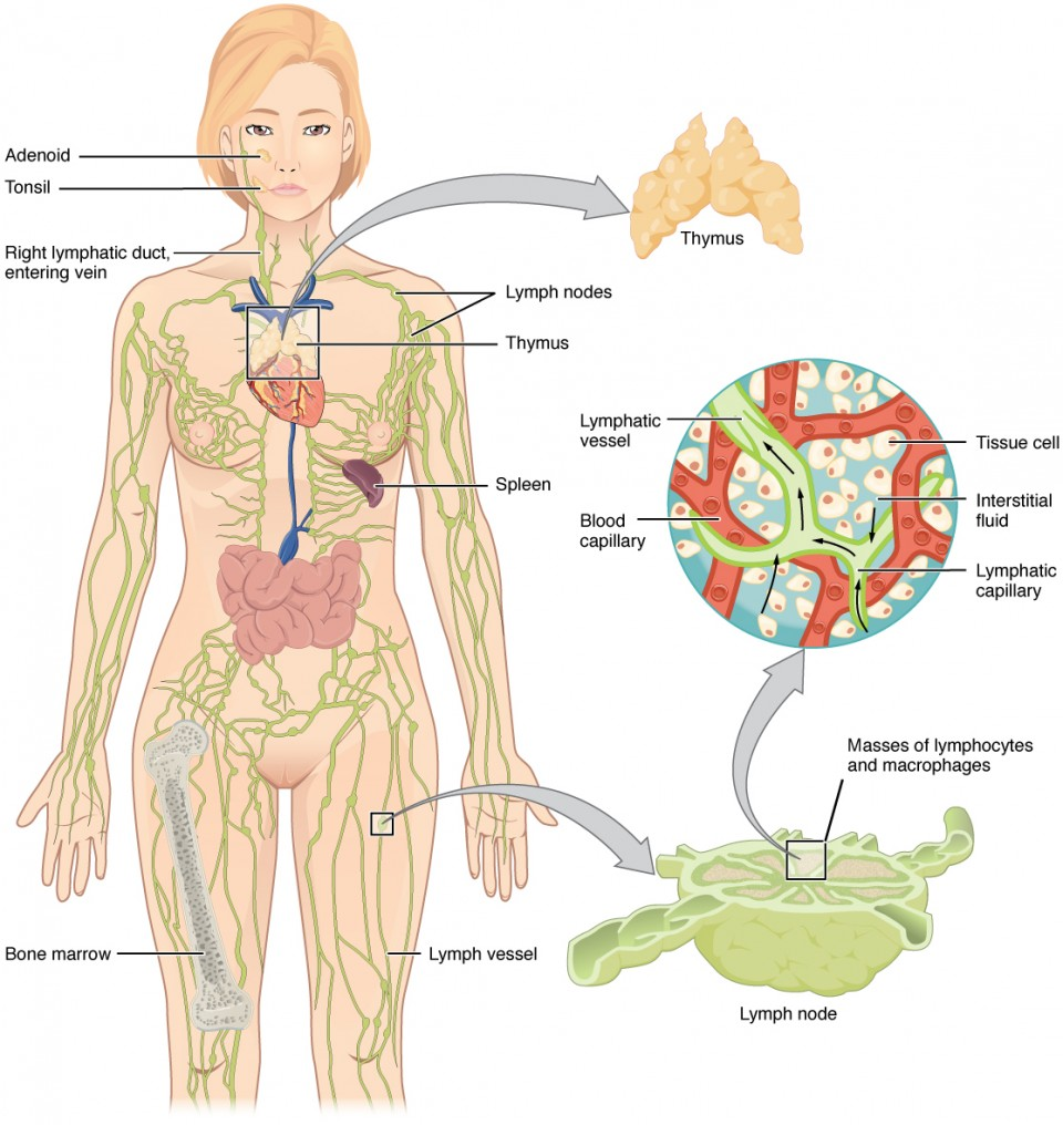
The lymphatic system is the system of vessels, cells, and organs that carries excess fluids to the bloodstream and filters pathogens from the blood.
Functions of the Lymphatic System
A major function of the lymphatic system is to drain body fluids and return them to the bloodstream.
Blood pressure causes leakage of fluid from the capillaries, resulting in the accumulation of fluid in the interstitial space—that is, spaces between individual cells in the tissues.
Lymph is the term used to describe interstitial fluid once it has entered the lymphatic system.
A lymph node is one of the small, bean-shaped organs located throughout the lymphatic system.
Lymphatic Capillaries
Lymphatic capillaries, also called the terminal lymphatics, are vessels where interstitial fluid enters the lymphatic system to become lymph fluid.
In the small intestine, dietary triglycerides combine with other lipids and proteins, and enter the lacteals to form a milky fluid called chyle.
Larger Lymphatic Vessels, Trunks, and Ducts
The superficial and deep lymphatics eventually merge to form larger lymphatic vessels known as lymphatic trunks.
The thoracic duct itself begins just beneath the diaphragm in the cisterna chyli, a sac-like chamber that receives lymph from the lower abdomen, pelvis, and lower limbs by way of the left and right lumbar trunks and the intestinal trunk.
The right lymphatic duct receives lymph from only the upper right side of the body.
The lymph from the rest of the body enters the bloodstream through the thoracic duct via all the remaining lymphatic trunks.
The Organization of Immune Function
Barrier defenses such as the skin and mucous membranes, which act instantaneously to prevent pathogenic invasion into the body tissue.
The slower but more specific and effective adaptive immune response, which involves many cell types and soluble factors, but is primarily controlled by white blood cells (leukocytes) known as lymphocytes, which help control immune responses
Lymphocytes: B Cells, T Cells, Plasma Cells, and Natural Killer Cells
B Cells
B cells are immune cells that function primarily by producing antibodies.
An antibody is any of the group of proteins that binds specifically to pathogen-associated molecules known as antigens.
An antigen is a chemical structure on the surface of a pathogen that binds to T or B lymphocyte antigen receptors.
T Cells: The T cell, on the other hand, does not secrete antibody but performs a variety of functions in the adaptive immune response.
Plasma Cells: A plasma cell is a B cell that has differentiated in response to antigen binding, and has thereby gained the ability to secrete soluble antibodies.
Natural Killer Cells: A natural killer cell (NK) is a circulating blood cell that contains cytotoxic (cell-killing) granules in its extensive cytoplasm.
Primary Lymphoid Organs and Lymphocyte Development
The primary lymphoid organs are the bone marrow, spleen, and thymus gland. The lymphoid organs are where lymphocytes mature, proliferate, and are selected, which enables them to attack pathogens without harming the cells of the body.
The red bone marrow is a loose collection of cells where hematopoiesis occurs, and the yellow bone marrow is a site of energy storage, which consists largely of fat cells.
The B cell undergoes nearly all of its development in the red bone marrow, whereas the immature T cell, called a thymocyte, leaves the bone marrow and matures largely in the thymus gland.
The thymus gland is a bilobed organ found in the space between the sternum and the aorta of the heart
Secondary Lymphoid Organs and their Roles in Active Immune Responses
Lymphocytes develop and mature in the primary lymphoid organs, but they mount immune responses from the secondary lymphoid organs.
A naïve lymphocyte is one that has left the primary organ and entered a secondary lymphoid organ.
Lymph nodes function to remove debris and pathogens from the lymph, and are thus sometimes referred to as the “filters of the lymph”.
In addition to the lymph nodes, the spleen is a major secondary lymphoid organ.
The other lymphoid tissues, the lymphoid nodules, have a simpler architecture than the spleen and lymph nodes in that they consist of a dense cluster of lymphocytes without a surrounding fibrous capsule.
Tonsils are lymphoid nodules located along the inner surface of the pharynx and are important in developing immunity to oral pathogens.
Mucosa-associated lymphoid tissue (MALT) consists of an aggregate of lymphoid follicles directly associated with the mucous membrane epithelia.
Bronchus-associated lymphoid tissue (BALT) consists of lymphoid follicular structures with an overlying epithelial layer found along the bifurcations of the bronchi, and between bronchi and arteries.
Barrier Defenses and the Innate Immune Response
The innate immune system enhances adaptive immune responses so they can be more effective.
Barrier Defenses
Site | Specific defense | Protective aspect |
|---|---|---|
Skin | Epidermal surface | Keratinized cells of surface, Langerhans cells |
Skin (sweat/secretions) | Sweat glands, sebaceous glands | Low pH, washing action |
Oral cavity | Salivary glands | Lysozyme |
Stomach | Gastrointestinal tract | Low pH |
Mucosal surfaces | Mucosal epithelium | Nonkeratinized epithelial cells |
Normal flora (nonpathogenicbacteria) | Mucosal tissues | Prevent pathogens from growing on mucosal surfaces |
A phagocyte is a cell that is able to surround and engulf a particle or cell, a process called phagocytosis.
A macrophage is an irregularly shaped phagocyte that is amoeboid in nature and is the most versatile of the phagocytes in the body.
A neutrophil is a phagocytic cell that is attracted via chemotaxis from the bloodstream to infected tissues.
A monocyte is a circulating precursor cell that differentiates into either a macrophage or dendritic cell, which can be rapidly attracted to areas of infection by signal molecules of inflammation.
Recognition of Pathogens
A pattern recognition receptor (PRR) is a membrane-bound receptor that recognizes characteristic features of a pathogen and molecules released by stressed or damaged cells.
A cytokine is signaling molecule that allows cells to communicate with each other over short distances.
A chemokine is a soluble chemical mediator similar to cytokines except that its function is to attract cells (chemotaxis) from longer distances.
Interferons are an example of early induced proteins.
Opsonization is the tagging of a pathogen for phagocytosis by the binding of an antibody or an antimicrobial protein.
Complement System
The complement system is a series of proteins constitutively found in the blood plasma.
Inflammatory Response
The hallmark of the innate immune response is inflammation.
Acute inflammation is a short-term inflammatory response to an insult to the body.
Chronic inflammation is ongoing inflammation.
There are four important parts to the inflammatory response:
Tissue Injury - The released contents of injured cells stimulate the release of mast cell granules and their potenT inflammatory mediators such as histamine, leukotrienes, and prostaglandins.
Vasodilation - Many inflammatory mediators such as histamine are vasodilators that increase the diameters of local capillaries.
Recruitment of Phagocytes - Leukotrienes are particularly good at attracting neutrophils from the blood to the site of infection by chemotaxis.
The Adaptive Immune Response: T lymphocytes and Their Functional Types
The immune system’s first exposure to a pathogen is called a primary adaptive response.
The secondary adaptive response often eliminates a pathogen before it can cause significant
tissue damage or any symptoms.
The basis of immunological memory, which protects us from getting diseases repeatedly from the same pathogen. By this mechanism, an individual’s exposure to pathogens early in life spares the person from these diseases later in life.
Antigens
Antigens on pathogens are usually large and complex, and consist of many antigenic determinants.
An antigenic determinant (epitope) is one of the small regions within an antigen to which a receptor can bind, and antigenic determinants are limited by the size of the receptor itself.
Antigen Processing and Presentation
Antigen processing is a mechanism that enzymatically cleaves the antigen into smaller pieces.
The antigen fragments are then brought to the cell’s surface and associated with a specialized type of antigen-presenting protein known as a major histocompatibility complex (MHC) molecule.
The association of the antigen fragments with an MHC molecule on the surface of a cell is known as antigen presentation and results in the recognition of antigen by a T cell.
T Cell Development and Differentiation
The process of eliminating T cells that might attack the cells of one’s own body is referred to as T cell tolerance.
In negative selection, self-antigens are brought into the thymus from other parts of the body by professional antigen-presenting cells.
Clonal selection is the process of antigen binding only to those T cells that have receptors specific to that antigen.
A clone is a group of lymphocytes that share the same antigen receptor.
A polyclonal response is the stimulation of multiple T cell clones.
Helper T Cells and their Cytokines
Helper T cells (Th), bearing the CD4 molecule, function by secreting cytokines that act to enhance other immune responses.
Th1 cells are a type of helper T cell that secretes cytokines that regulate the immunological activity and development of a variety of cells, including macrophages and other types of T cells.
Th2 cells, on the other hand, are cytokine-secreting cells that act on B cells to drive their differentiation into plasma cells that make antibody.
Cytotoxic T cells (Tc) are T cells that kill target cells by inducing apoptosis using the same mechanism as NK cells.
The Adaptive Immune Response: B-lymphocytes and Antibodies
Scientists now know the cause of the agglutination is an antibody molecule, also called an immunoglobulin.
Central tolerance is the destruction or inactivation of B cells that recognize self-antigens in the bone marrow, and its role is critical and well established.
In the process of clonal deletion, immature B cells that bind strongly to self-antigens expressed on tissues are signaled to commit suicide by apoptosis, removing them from the population.
In peripheral tolerance, functional, mature B cells leave the bone marrow but have yet to be exposed to self-antigen.
The heavy chain and the light chain are the two polypeptides that form the antibody.
The Fc region of the antibody is formed by the two heavy chains coming together, usually linked by disulfide bonds.
Five Classes of Antibodies and their Functions
IgM consists of five four-chain structures (20 total chains with 10 identical antigen-binding sites) and is thus the largest of the antibody molecules.
IgG is a major antibody of late primary responses and the main antibody of secondary responses in the blood.
IgA exists in two forms, a four-chain monomer in the blood and an eight-chain structure, or dimer, in exocrine gland secretions of the mucous membranes, including mucus, saliva, and tears.
IgE is usually associated with allergies and anaphylaxis.
Active versus Passive Immunity
Active immunity is the resistance to pathogens acquired during an adaptive immune response within an individual.
Passive immunity arises from the transfer of antibodies to an individual without requiring them to mount their own active immune response.
T cell-dependent versus T cell-independent Antigens
A T cell-independent antigen usually is in the form of repeated carbohydrate moieties found on the cell walls of bacteria.
A T cell-dependent antigen, on the other hand, usually is not repeated to the same degree on the pathogen and thus does not crosslink surface antibody with the same efficiency.
The Immune Response against Pathogens
Seroconversion is the reciprocal relationship between virus levels in the blood and antibody levels.
Neutralization is the process of coating a pathogen with antibodies, making it physically impossible for the pathogen to bind to receptors.
Macrophage oxidative metabolism is hostile to intracellular bacteria, often relying on the production of nitric oxide to kill the bacteria inside the macrophage.
Autoimmune Responses
Disease | Autoantigen | Symptoms |
|---|---|---|
Celiac disease | Tissue transglutaminase | Damage to small intestine |
Diabetes mellitus type I | Beta cells of pancreas | Low insulin production; inability to |
Graves’ disease | Thyroid-stimulating hormone receptor (antibody blocksreceptor) | Hyperthyroidism |
Transplantation and Cancer Immunology
Tissue typing is the determination of MHC molecules in the tissue to be transplanted to better match the donor to the recipient.
An interesting consequence of Rh factor expression is seen in erythroblastosis fetalis, a hemolytic disease of the newborn.
MHC polygeny refers to the multiple MHC proteins on cells, and MHC polymorphism refers to the multiple alleles for each individual MHC locus.