


Chapter 14: The Somatic Nervous System

Sensory Receptors
Sensory receptors are specialized cells that detect changes in the environment and convert them into electrical signals that can be interpreted by the brain.
They are found in the skin, eyes, ears, nose, and other organs, and are responsible for detecting sensations such as touch, pressure, temperature, sound, and light.
Structural Receptor Types
The cells in the retina that respond to light stimuli are an example of a specialized receptor, a photoreceptor.
An exteroceptor is a receptor that is located near a stimulus in the external environment, such as the somatosensory receptors that are located in the skin.
An interoceptor is one that interprets stimuli from internal organs and tissues, such as the receptors that sense the increase in blood pressure in the aorta or carotid sinus.
A proprioceptor is a receptor located near a moving part of the body, such as a muscle, that interprets the positions of the tissues as they move.
Functional Receptor Types
Chemical stimuli can be interpreted by a chemoreceptor that interprets chemical stimuli, such as an object’s taste or smell.
Osmoreceptors respond to solute concentrations of body fluids.
Additionally, pain is primarily a chemical sense that interprets the presence of chemicals from tissue damage, or similar intense stimuli, through a nociceptor.
Physical stimuli, such as pressure and vibration, as well as the sensation of sound and body position (balance), are interpreted through a mechanoreceptor.
Sensory Modalities
A general sense is one that is distributed throughout the body and has receptor cells within the structures of other organs.
General senses often contribute to the sense of touch, or to proprioception (body movement) and kinesthesia (body movement), or to a visceral sense, which is most important to autonomic functions.
A special sense is one that has a specific organ devoted to it, namely the eye, inner ear, tongue, or nose.
The general sense of touch, which is known as somatosensation, can be separated into light pressure, deep pressure, vibration, itch, pain, temperature, or hair movement.
Gustation (Taste)
Only a few recognized submodalities exist within the sense of taste, or gustation.
Raised bumps called papillae (singular = papilla) contain the structures for gustatory transduction.
Within the structure of the papillae are taste buds that contain specialized gustatory receptor cells for the transduction of taste stimuli.
Alkaloids are nitrogen-containing molecules that often have a basic pH.
Olfaction (Smell)
The olfactory receptor neurons are located in a small region within the superior nasal cavity. This region is referred to as the olfactory epithelium and contains bipolar sensory neurons.
Each olfactory sensory neuron has dendrites that extend from the apical surface of the
epithelium into the mucus lining the cavity.
These odorant molecules bind to proteins that keep them dissolved in the mucus and help transport them to the olfactory dendrites.
The group of axons called the olfactory tract connect to the olfactory bulb on the ventral surface of the frontal lobe.
Audition (Hearing)
Hearing, or audition, is the transduction of sound waves into a neural signal that is made possible by the structures of the ear.
The large, fleshy structure on the lateral aspect of the head is known as the auricle.
At the end of the auditory canal is the tympanic membrane, or ear drum, which vibrates after it is struck by sound waves.
The auricle, ear canal, and tympanic membrane are often referred to as the external ear.
The middle ear consists of a space spanned by three small bones called the ossicles.
The three ossicles are the malleus, incus, and stapes, which are Latin names that roughly translate to hammer, anvil, and stirrup.
The stapes is then attached to the inner ear, where the sound waves will be transduced into a neural signal.
Sound is transduced into neural signals within the cochlear region of the inner ear, which contains the sensory neurons of the spiral ganglia.
The cochlea is attached to the stapes through the oval window.
The oval window is located at the beginning of a fluid-filled tube within the cochlea called the scala vestibuli.
The scala tympani ends at the round window, which is covered by a membrane that contains the fluid within the scala.
Equilibrium (Balance)
Along with audition, the inner ear is responsible for encoding information about equilibrium, the sense of balance.
Head position is sensed by the utricle and saccule, whereas head movement is sensed by the semicircular canals.
The base of each semicircular canal, where it meets with the vestibule, connects to an enlarged region known as the ampulla.
The stereocilia of these hair cells extend into the cupula, a membrane that attaches to the top of the ampulla.
Somatosensation (Touch)
Somatosensation (Touch) is the sense of touch, pressure, temperature, and pain that is detected by the skin and other body parts.
It is the ability to detect and interpret physical sensations from the environment.
Vision
Vision is the special sense of sight that is based on the transduction of light stimuli received through the eyes.
The inner surface of each lid is a thin membrane known as the palpebral conjunctiva.
Tears are produced by the lacrimal gland, located beneath the lateral edges of the nose.
Movement of the eye within the orbit is accomplished by the contraction of six extraocular muscles that originate from the bones of the orbit and insert into the surface of the eyeball.
The superior oblique originates at the posterior orbit, near the origin of the four rectus muscles.
The inferior oblique muscle originates from the floor of the orbit and inserts into the inferolateral surface of the eye.
A seventh muscle in the orbit is the levator palpebrae superioris, which is responsible for elevating and retracting the upper eyelid, a movement that usually occurs in concert with elevation of the eye by the superior rectus.
The middle layer of the eye is the vascular tunic, which is mostly composed of the choroid, ciliary body, and iris.
The choroid is a layer of highly vascularized connective tissue that provides a blood supply to the eyeball.
Overlaying the ciliary body, and visible in the anterior eye, is the iris—the colored part of the eye.
The innermost layer of the eye is the neural tunic, or retina, which contains the nervous tissue responsible for photoreception.
The anterior cavity is the space between the cornea and lens, including the iris and ciliary body. It is filled with a watery fluid called the aqueous humor.
The posterior cavity is filled with a more viscous fluid called the vitreous humor.
At the exact center of the retina is a small area known as the fovea.
Visual acuity, or the sharpness of vision, is greatest at the fovea.
Photoreceptor cells have two parts, the inner segment and the outer segment segment. The rod-shaped outer segments of the rod photoreceptor contain a stack of membrane-bound discs that contain the photosensitive pigment rhodopsin.
The cone-shaped outer segments of the cone photoreceptor contain their photosensitive pigments in infoldings of the cell membrane.
There are three cone photopigments, called opsins, which are each sensitive to a particular wavelength of light.
A single unit of light is called a photon, which is described in physics as a packet of energy with properties of both a particle and a wave.
Opsin pigments are actually transmembrane proteins that contain a cofactor known as retinal.
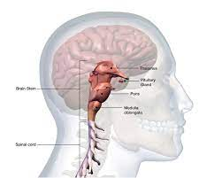
Spinal Cord and Brain Stem
A sensory pathway that carries peripheral sensations to the brain is referred to as an ascending pathway, or ascending tract.
The dorsal column system (sometimes referred to as the dorsal column–medial lemniscus) and the spinothalamic tract are two major pathways that bring sensory information to the brain
The dorsal column is separated into two component tracts, the fasciculus gracilis that contains axons from the legs and lower body, and the fasciculus cuneatus that contains axons from the upper body and arms.
The nucleus gracilis is the target of fibers in the fasciculus gracilis, whereas the nucleus cuneatus is the target of fibers in the fasciculus cuneatus.
These axons then continue to ascend the brain stem as a bundle called the medial lemniscus.
The spinal trigeminal nucleus of the medulla receives information similar to that carried by spinothalamic tract, such as pain and temperature sensations.
Other axons go to either the chief sensory nucleus in the pons or the mesencephalic nuclei in the midbrain.
The sensory pathway for gustation travels along the facial and glossopharyngeal cranial nerves, which synapse with neurons of the solitary nucleus in the brain stem.
Axons from the solitary nucleus then project to the ventral posterior nucleus of the thalamus.
Auditory processing continues on to a nucleus in the midbrain called the inferior colliculus.
The majority of the connections of the optic tract are to the thalamus—specifically, the lateral geniculate nucleus.
Cortical Responses
The most anterior regions of the frontal lobe—the prefrontal areas—are important for executive functions, which are those cognitive functions that lead to goal-directed behaviors.
These higher cognitive processes include working memory, which has been called a “mental scratch pad,” that can help organize and represent information that is not in the immediate environment.
The prefrontal areas project into the secondary motor cortices, which include the premotor cortex and the supplemental motor area.
The frontal eye fields are responsible for moving the eyes in response to visual stimuli.
Anterior to the premotor cortex and primary motor cortex is Broca’s area.
It then passes between the caudate nucleus and putamen of the basal nuclei as a bundle called the internal capsule.
The tract then passes through the midbrain as the cerebral peduncles, after which it burrows through the pons.
The lateral corticospinal tract is composed of the fibers that cross the midline at the pyramidal decussation.
The cervical enlargement is particularly large because there is greater control over the fine musculature of the upper limbs, particularly of the fingers.
The lumbar enlargement is not as significant in appearance because there is less fine motor control of the lower limbs.
The anterior corticospinal tract is responsible for controlling the muscles of the body trunk
Other descending connections between the brain and the spinal cord are called the extrapyramidal system.
The reticulospinal tract connects the reticular system, a diffuse region of gray matter in the brain stem, with the spinal cord.
The vestibulospinal tract connects the brain stem nuclei of the vestibular system with the spinal cord.